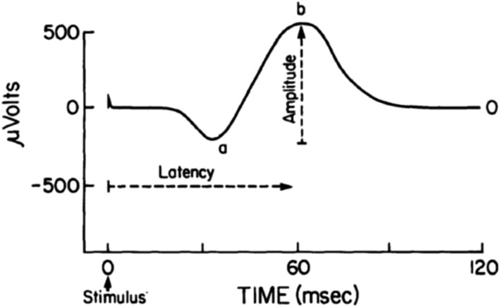
FIGURE 8.
The electroretinogram response in the μvolts is generated by light stimulation of the retina and occurs in milliseconds. The electroretinogram is characterized by a negative “a” wave originating from the photoreceptor inner segments followed by a positive “b” wave, which originates from the retinal bipolar cell layer. The electroretinogram amplitude is proportional to the intensity of light, the degree of dark adaptation, the number of photoreceptors, and the rhodopsin visual pigment concentration. Reproduced from reference 204 with permission.

