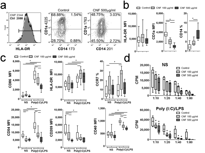
Figure 2. Effects of CNFs on differentiation, Poly (I:C)/LPS-induced maturation and allostimulatory capacity of mo-DCs.
(a) The expression of HLA-DR, CD1a and CD14 was analyzed on immature DCs generated from monocytes in GM-CSF/IL-4-supplemented medium in the presence or absence of CNFs (Control) after 5 days. The results from one representative experiment, or (b) 8 experiments (mo-DC donors) are shown. (c) The phenotypic analysis of mo-DCs which were stimulated with Poly (I:C)/LPS, or were left non-stimulated (NS) for additional 2 days was carried out, and the results from 9 experiments are shown. (d) The proliferation in co-culture with either NS or Poly (I:C)/LPS-matured mo-DCs, and MACS purified CD4+T cells (1 × 10⁵ cells/well), carried out in different mo-DC-to-T cell ratios (1:10–1:80), was measured by 3H-thymidin incorporation assay after 5 days (cpm, counts per minute). The results from 8 different mo-DC/CD4 proliferation assays are shown. *p < 0.05 compared to control, or as indicated by line (Friedman test with Dunns posttest) NS - non-stimulated.

