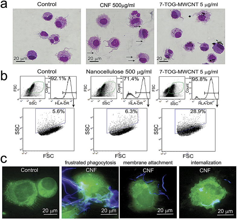
Figure 5. Internalization of CNFs by mo-DCs.
(a) Mo-DCs differentiated in the presence or absence of CNFs were collected and the cytospins were stained with MGG. Immature control mo-DCs were additionally cultivated with 7-TOG-MWCNT, which was used as a positive control. Arrows mark the grayish CNFs on the samples. (b) The same samples were analyzed by flow cytometry after labeling of mo-DCs with anti-HLA-DR-Alexa 488 Ab. (c) Cytospins of mo-DCs cultivated with CNFs were stained in anti-HLA-DR-Alexa 488 and Calcofluor white, and then analyzed by the epi-fluorescent microscopy. Representative data and images are shown, collected from 5 different experiments.

