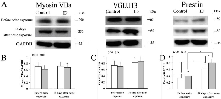
Figure 6.
Expression of myosin VIIa, vesicular glutamate transporter (VGLUT3) and prestin in the cochleae before noise exposure (on PND 21) and at 14 days after noise exposure (on PND 36). Representative immunoblots of myosin VIIa, VGLUT3 and prestin of young rat cochleae in control (left ear, n = 3) and ID group (left ear, n = 3) at respective time point (A); GAPDH was used as a loading control. Densitometry of immunoblots of myosin VIIa (B); VGLUT3 (C); and prestin (D) were compared, respectively, between two groups. The height of each bar represents the mean ± SEM (* p < 0.05).

