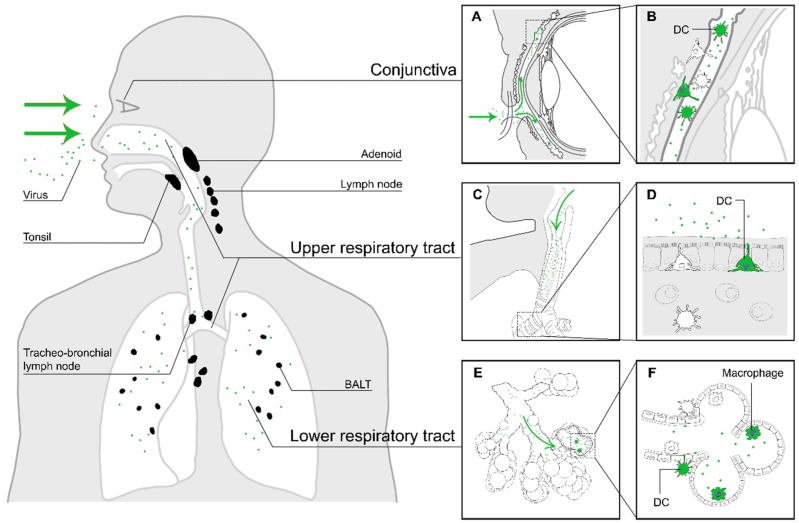
Figure 1.
The first stage of MV infection: entry of MV into a susceptible host. The virus enters the respiratory tract (green arrows in panels (C) and (E)), where it binds to DC-SIGN+ DCs or infects CD150+ myeloid or lymphoid cells in the mucocilliary epithelium or the alveolar spaces. Another potential site of entry is through the conjunctiva, which is rich in DCs and CD150+ lymphocytes (A). Panels on the right show an enlarged illustration of potential entry events. MV particles deposited on the conjunctiva will enter the space between cornea and eyelids ((A), green arrows), where they can infect myeloid or lymphoid cells (B). MV particles inhaled into the respiratory tract ((C) and (E), green arrows) can either infect DC-SIGN+ dendritic cells in the upper respiratory tract, with dendrites protruding into the respiratory mucosa (D), or dendritic cells or macrophages in the alveolar lumina of the lower respiratory tract (F). The infected immune cells subsequently migrate to nearby tertiary lymphoid tissues and draining lymph nodes (black).

