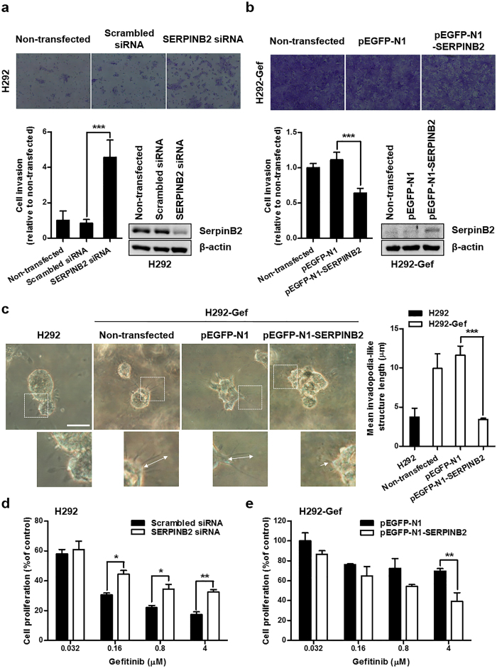
Figure 3. SerpinB2-dependent changes in invasive characteristics and gefitinib sensitivity in H292 and H292-Gef cells.
(a,b) H292 (a) and H292-Gef (b) cells were not transfected or transfected with indicated siRNAs or plasmids for 24 h, and altered gene expression was confirmed by western blot using a SerpinB2 antibody. The cell invasion assay was performed with transfected cells as described in the Methods section. Magnification, x100. The data are presented as the mean ± SD. (c) Cells were embedded into 3D collagen I gels and incubated for 2 days. On Day 2, cells were visualized by light microscopy and length of invadopodia-like structures was measured. The data are presented as the mean ± SD. Scale bar, 25 μm. (d,e) H292 (d) and H292-Gef (e) cells were transfected with indicated siRNAs or plasmids for 24 h. The transfected cells were plated in 96-well culture plates for 24 h and then treated with gefitinib for 48 h. The data are presented as the mean ± SD. *P < 0.05, **P < 0.01, ***P < 0.005.

