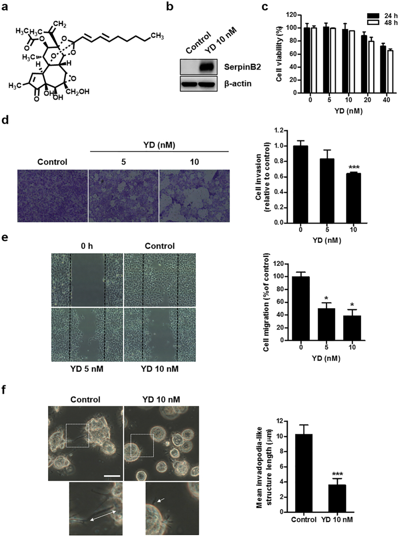
Figure 4. Effects of YD on the invasion and invadopodia-like structures in H292-Gef cells.
(a) Structure of yuanhuadine (YD). (b) H292-Gef cells were treated with 10 nM YD for 24 h and then lysed and subjected to western blot using anti-SerpinB2 and β-actin as an internal standard. (c) H292-Gef cells were treated with indicated concentrations of YD for 24 and 48 h, and cell viability was then determined using the MTT assay. The data are presented as the mean ± SD. (d) H292-Gef cells were added to Matrigel-coated transwell inserts with YD and incubated for 48 h. The invaded cells were fixed, stained and counted as described in the Methods section. Magnification, x100. The data are presented as the mean ± SD. (e) After mechanistically generating scratches in the monolayer of H292-Gef cells, cells were treated with YD for 30 h, and wound closure was observed under a light microscope. The percentage of cell migration was calculated as described in the Methods section. The data are presented as the mean ± SD. Magnification, x40. (f) H292-Gef cells embedded in 3D collagen I gels were treated with 10 nM YD for 24 h. Cells were visualized by light microscopy and length of invadopodia-like structures was measured. The data are presented as the mean ± SD. Scale bar, 25 μm. *P < 0.05, **P < 0.01, ***P < 0.005.

