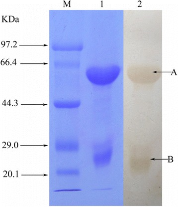
Fig. 3.

Identification of anti-T. albolabris venom IgY. The reduced IgY (50 μg) was separated on 10 % SDS-PAGE and stained with Coomassie brilliant blue (lane M: molecular weight marker; lane 1: reduced IgY). Result of Western blot test using peroxidase conjugated rabbit anti-chicken IgY to detect the heavy chains (lane 2: A) and the light chains (lane 2: B)
