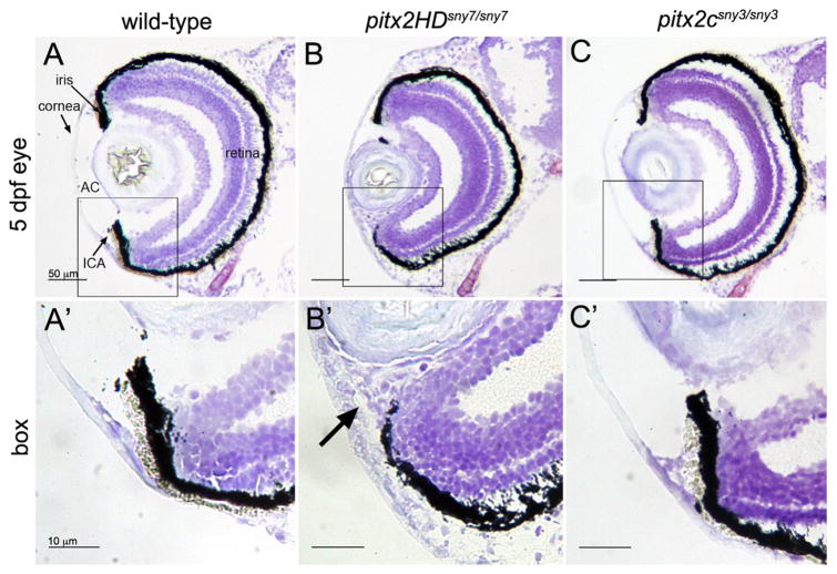
Fig. 4.
Malformations of the anterior segment of the eye in Pitx2HD mutant embryos. (A–C) Cryosections of the eye from 5 dpf wild-type (A), pitx2HDsny7/sny7 (B) and pitx2csny3/sny3 (C) fish stained with crystal violet. AC=anterior chamber; ICA=iridocorneal angle. Scale bars=50 μm. (A′–C′) Enlarged view of box 1 in A–C showing anterior segment structures. In pitx2HD mutants (B′), the anterior chamber was reduced and the iridocorneal angle (arrow) was malformed. Scale bars=10 μm.

