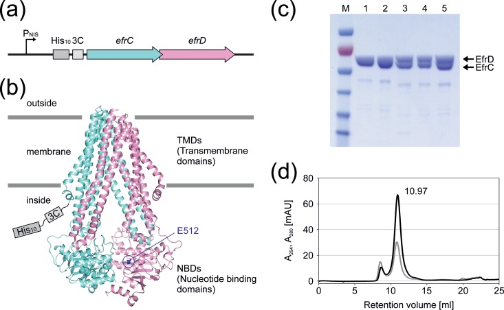
FIG 4.
Expression and purification of EfrCD. (a) L. lactis expression construct containing the deca-His tag and 3C protease cleavage site followed by the ORFs encoding EfrC and EfrD. (b) Homology model of EfrCD based on the coordinates of TM287/288. EfrC is shown in aquamarine and EfrD in pink, and the conserved Walker B glutamate (E512) of the consensus site is highlighted as blue sticks. The deca-His tag and 3C protease cleavage site are attached to the N terminus of EfrC. (c) SDS-PAGE analysis of the different purification steps. Lanes: 1, Ni2+-NTA elution; 2, PD-10 elution; 3, after 3C protease cleavage; 4, after Ni2+-NTA rebinding; 5, main peak fraction of size exclusion chromatography separation. (d) Size exclusion chromatogram of EfrCD using a Superdex 200 Increase 10/300 GL column. A280 is shown in black and A254 in gray. The main peak, eluting at ca. 11 ml, corresponds to an EfrCD heterodimer.

