FIG 1.
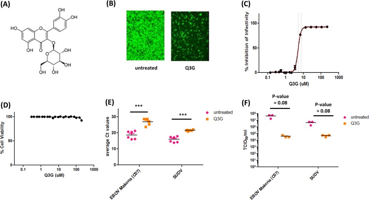
Q3G inhibits replication of Ebola virus in vitro. (A) Chemical structure of Q3G. (B) Vero E6 cells were pretreated with 10 μM Q3G for 1 h and then infected with EBOV-Kikwit-GFP (MOI = 0.1) at 37°C and incubated for 72 h in the presence of 10 μM Q3G. (C) The EC50 of Q3G against EBOV-Kikwit-GFP was determined by pretreating Vero E6 cells with serial dilutions of Q3G for 1 h and then infecting them with virus (MOI = 0.1) at 37°C for 72 h in the presence of Q3G, at which time fluorescence was quantified. (D) To determine cell viability following Q3G treatment, Vero E6 cells were treated with serial dilutions of Q3G. Viability was measured after 72 h by a resazurin dye-based assay, and data were normalized to data for untreated controls. (E) Inhibition of EBOV-Makona and SUDV was determined by pretreating cells with 10 μM Q3G for 1 h and then infecting them with virus (MOI = 0.1) at 37°C for 1 h. Cells were collected, and the amount of viral RNA was quantified at 5 days postinfection by RT-qPCR. Ct, threshold cycle. (F) Inhibition of EBOV-Makona and SUDV was determined by pretreating cells with 10 μM Q3G for 1 h and then infecting them with virus (MOI = 0.1) at 37°C for 1 h. Cells were collected, and the amount of viral RNA was quantified at 5 days postinfection by using a TCID50 assay. All experiments were performed in triplicate, and error bars represent the standard errors of the means. ***, P value of <0.001.

