
Fig. (6).
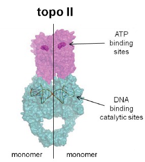
Model for the eukaryotic Type II topoisomerase, topo II, in covalent (“cleavable”) complex with DNA. This representation is a composite of the two PDB entries 1ZXN (N terminal human topo IIα with ATP analog ADPNP in the ATP binding site) from Wei et al. [133] and 3QX3 (C-terminal catalytic domain of human topo IIβ with bound and cleaved DNA and bound etoposide) from Wu et al. [134]. The pink represents the ATP binding domains, analogous to bacterial GyrB (or ParE) and the green represents the DNA binding domains, analogous to bacterial GyrA (or ParC). The vertical line indicates the enzyme dimeric composition as two symmetrical monomers (right and left).