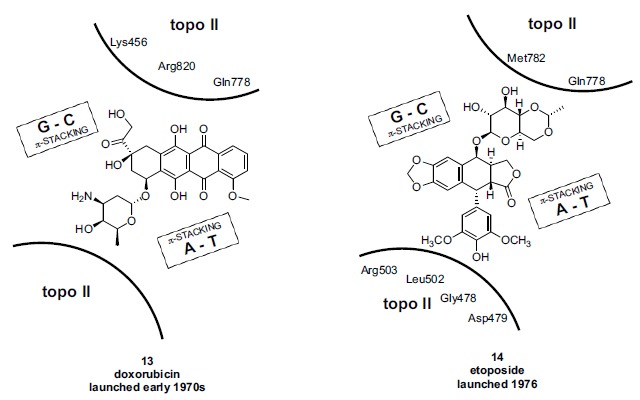
Fig. (7).

Schematic representation of anticancer agents doxorubicin 13 (left panel) and etoposide 14 (right panel) bound as the ternary complex (“cleavable complex”) with eukaryotic topo II and DNA. The binding modes of 13 and 14 are broadly similar to that of moxifloxacin 12 in the prokaryotic ternary complex (compare Fig. 5). Renderings adapted from crystal structure information by Wu et al. and Chan et al. [135, 136] employing topo IIβ. Topo II amino acids which form known interactions with the drugs based on crystallography are shown in each of the two panels, although the specific interactions are not depicted. A strand break between two base pairs, mediated by a topoisomerase catalytic tyrosine, is not shown.
