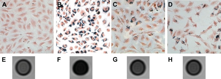
Figure 8.
In vitro labeling of TVECs with CL-PEG-MnFe2O4.
Notes: Prussian blue staining (200×) of control group (A), TVECs labeled with CL-PEG-MnFe2O4 (B), PEG-b-PCL-MnFe2O4 (C) and the mixture of CL-PEG-MnFe2O4 and CL 1555 peptides (D). It showed that the uptake of CL-PEG-MnFe2O4 was high, while few PEG-b-PCL-MnFe2O4 was engulfed into TVECs. Blocking CD105 with the free CL 1555 peptides also effectively reduced the amount of blue granules in the cytoplasm of TVECs. (E–H) showed the T2-weighted MR images of the four groups of cells. Correspondingly, the cells co-cultured with CL-PEG-MnFe2O4 (F) showed a more noticeable signal intensity drop than that with PEG-b-PCL-MnFe2O4 (G) and blocking with the free CL 1555 peptides (H).
Abbreviations: MR, magnetic resonance; PEG-b-PCL, polyethylene glycol-block-poly(ε-caprolactone); TVECs, tumor vascular endothelial cells.

