Figure 5.
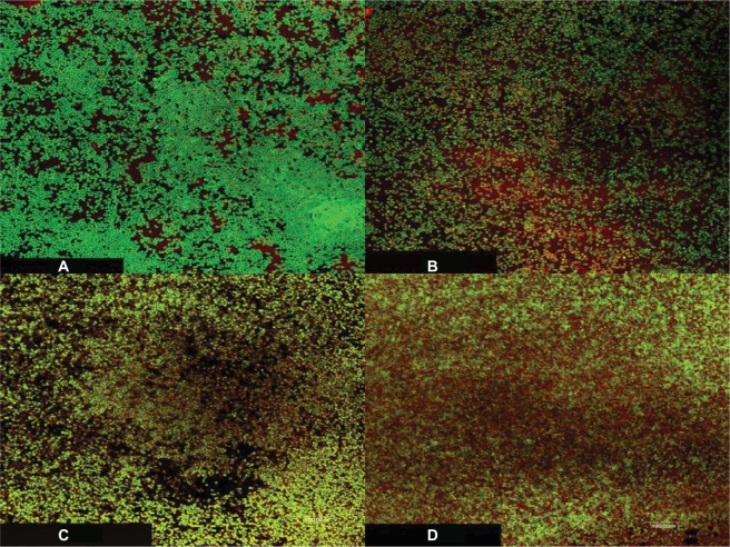
CLSM images of S. epidermidis biofilms on plastic coverslips after incubation for 48 hours followed by one of the following treatments: (A) control (untreated), (B) UVC light alone followed by 24 hours of incubation, (C) VAN alone at twice of its MBC and incubation for 24 hours, and (D) UVC light followed by VAN at double of its MBC and incubation for 24 hours.
Notes: The bacterial cells were stained with LIVE/DEAD BacLight bacterial viability stain to directly visualize the effects of the UVC light and the antibiotic. The green fluorescence reflects processing of the dye by metabolically active cells, while the red fluorescence is characteristic of dead cells. The green fluorescence was considerably prominent in all the samples with few dead cells when the biofilm was treated with either the UVC light or the antibiotic alone, and the dead cells increased when both were used in sequence. Magnification 1000×.
Abbreviations: CLSM, confocal laser scanning microscopy; S. epidermidis, Staphylococcus epidermidis; UVC, ultraviolet C; VAN, vancomycin; MBC, minimum bactericidal concentration.

