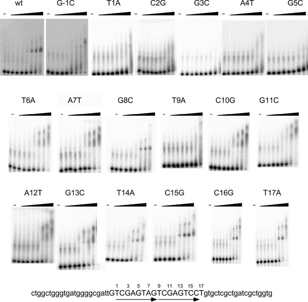
Figure 5.
Mutational analysis of parS. The substrate for all EMSAs shown here is at the bottom of the figure. It contains the 18 bp predicted parS site and is flanked by nonspecific DNA. This sequence contains two copies of a tandem repeated sequence (shown by arrows). ParB binds to this substrate, but when single base mutations are made and binding is assayed, it is clear that the first repeat sequence is more important for binding. ParB protein concentrations are the same as in Figure 4.

