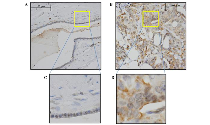
Figure 1.
Representative KIF18A immunohistochemistry images of breast cancer tissues. Positive staining was observed in cancer cells, but very limited in normal cells. (A) Normal breast, magnification ×400. (B) Breast cancer, magnification ×400. Enlarged views of (C) normal breast and (D) breast cancer tissues, magnification ×1,000.

