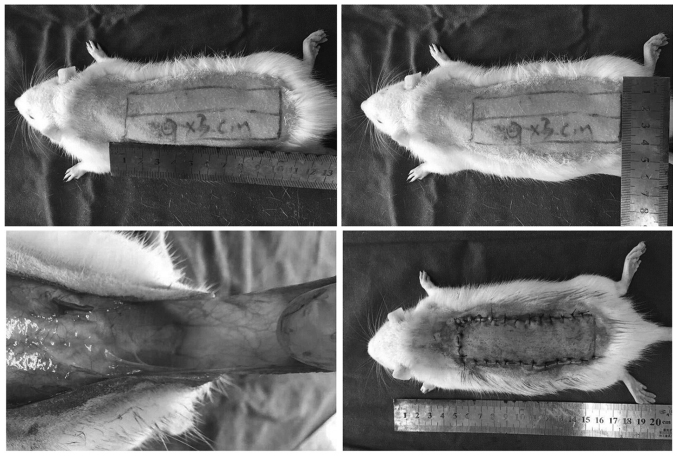
Figure 1.
A McFarlane flap model was designed (size, 9×3 cm) on the back of each rat and both sacral arteries were systematically sectioned, so that no axial vessels were incorporated into the flap. The flap was separated from the underlying fascia up to its base. After controlling any bleeding, the flap was immediately sutured to the original position with 4-0 running nylon sutures and a wedged cutting needle.

