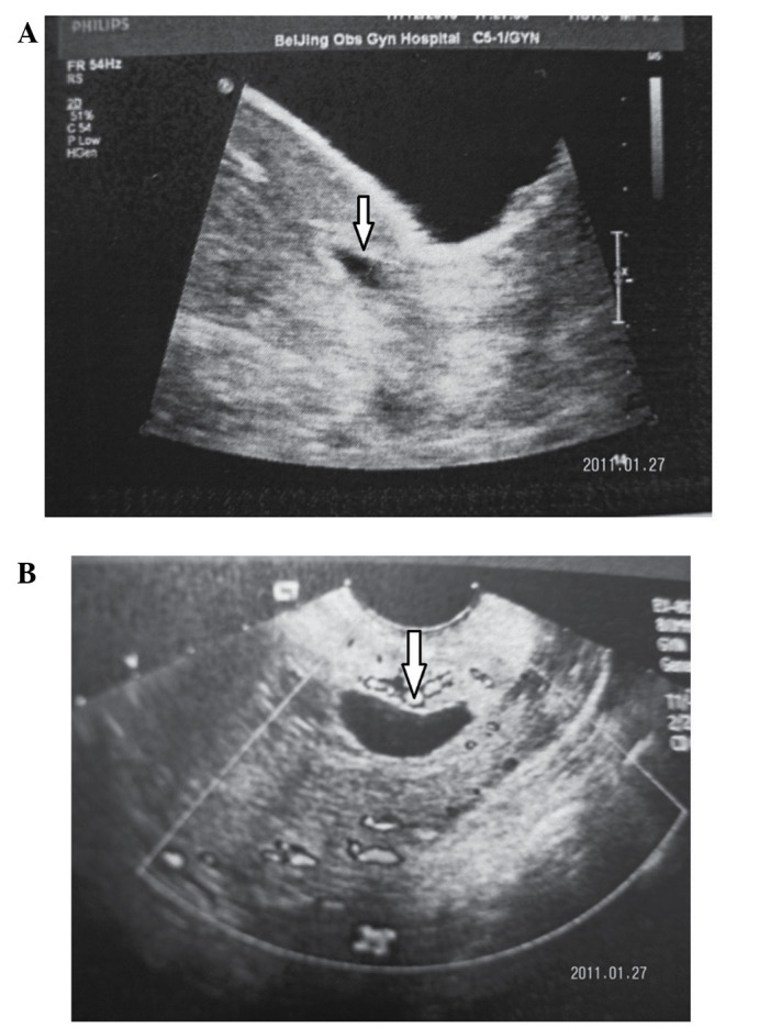
Figure 1.

Two examples of echo-images of CSP. Images were captured from a 30-year-old woman at 6 weeks of CSP with a history of one caesarean delivery. (A) TAS showing the midline of the uterus. (B) Transverse TVS showing the midline of the uterus. Arrow shows pregnant scar. CSP, caesarean scar pregnancy; TAS, transabdominal sonography; TVS, transvaginal sonography.
