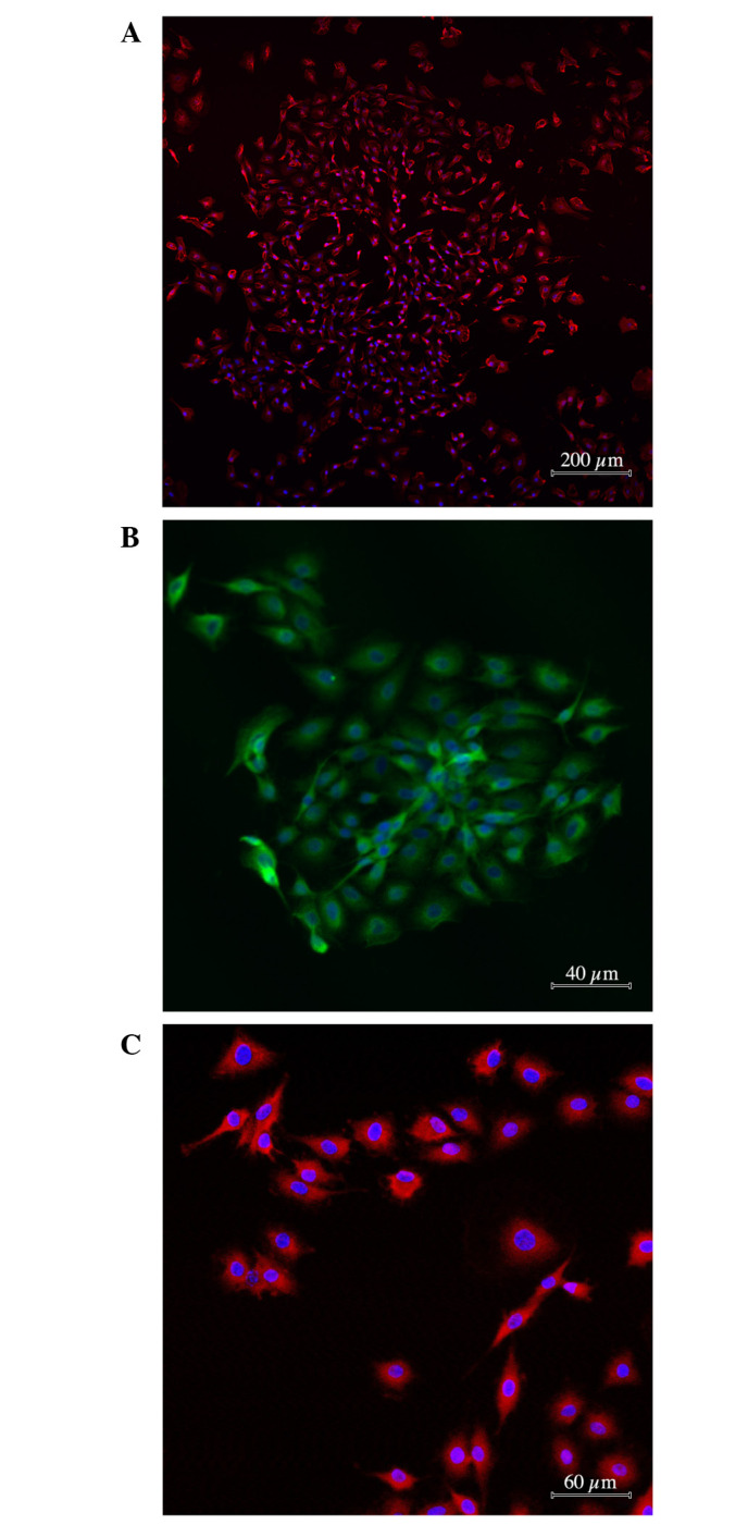
Figure 1.

Immunocytochemical characteristic of glioma cells used in the experiment. Antibody staining against (А) nestin, (B) p53 and (C) C-X-C chemokine receptor type 4. Cytoplasms are stained with (A and C) Alexa 633 (pink) or (B) Alexa 488 (green). Nuclei are counterstained by DAPI (blue).
