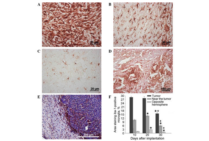
Figure 4.
Tumor in the rat brain. Immunocytochemical antibody staining for IBA-1 (microglia/macrophage-specific protein) in (A) the neoplastic nodule, (B) the brain area adjacent to the nodule and (C) the brain area of the hemisphere opposite to the nodule 20 days after implantation, and in (D) the neoplastic nodule 30 days after implantation. (E) Staining with hematoxylin and eosin and IBA-1 antibody revealed IBA-positive cells on the border of the neoplastic nodule on day 30 after implantation. (F) IBA-1-positive cell performance dynamics in neoplastic nodule over time *P<0.05 vs. 10 days; +P<0.05 vs. 20 days. IBA-1, ionized calcium-binding adapter molecule-1.

