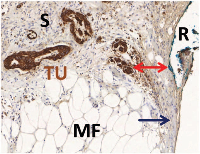
FIGURE 1.

Conventional and histopathological margin status assessment. Example of a tissue slide from the mesopancreatic margin with brown immunohistochemical staining for Pan-Cytokeratin for better visualization of tumor cells. The tumor cells (TU) are surrounded by a dense fibrotic stroma (S) and invade the mesopancreatic fatty tissue (MF). The closest distance to the inked resection margin (R) is marked by a red arrow. Although no tumor cells are found directly at the resection margin, there is broad contact of the fibrotic stroma to the resection margin. Margin status in this case is negative by conventional R-status (R0, zero tumor cell distance rule), but positive by circumferential margin concept (CRM+, 1-mm tumor cell distance rule) and positive by stromal clearance concept (S+, zero stroma distance rule). For details see manuscript text.
