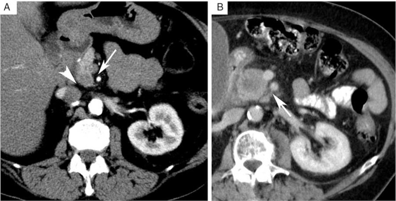
FIGURE 2.

Assessment of radiologic parameters. A, Preoperative contrast enhanced multiphasic multidetector computed tomography (MDCT) demonstrating a normal fatty tissue sheath separating the superior mesenteric artery (SMA, arrow) and the inferior caval vein (ICV, arrowhead) from the pancreas. A small hypovascular/hypodense tumor in the pancreatic head can be seen. B, Preoperative contrast enhanced MDCT demonstrating the presence of stranding, that is increased attenuation, in the SMA fatty tissue sheath (arrow).
