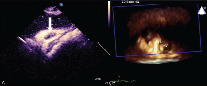
FIGURE 2.

Contrast enhancement ultrasound findings show few contrast agent enhancements in the tumor structure (white arrow; A); real-time 3D full-volume bird's eye view findings of the mass adherent to the IVS and attached to the RV wall (B). IVS = interventricular septum; MV = bicuspid valve; RV = right ventricle.
