
FIGURE 10.
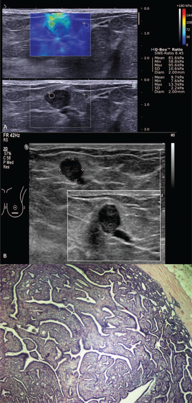
Shearwave elastography (SWE) of a Breast Imaging-Reporting and Data System (BI-RADS) 4a lesion in a 27-year-old woman with a palpable lump and nipple discharge. (A) Shearwave imaging showed a heterogeneous color map and Emax of 95.6 kPa. (B) Gray scale ultrasound showed a lobulated hypoechoic lesion continuous with a dilated duct. (C) Photomicrograph (H and E, ×40) of surgical excision specimen of an intraductal papilloma.