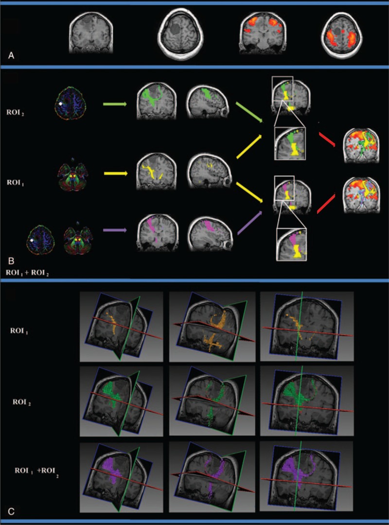
FIGURE 4.

The CST fiber tracking results for patient 2, who was diagnosed with transitional meningioma in the right frontal cortex that did not affect the primary motor cortex or the right internal capsule. (A) The lesion and motor-activated area are displayed on a T1-weighted image. (B) The CST fiber tracking results based on different ROI definitions. Yellow, green, and purple represent the CST fiber tracking results obtained using ROI1, ROI2, and ROI1 + ROI2, respectively. fMRI activation is observed in the primary and supplementary motor cortex. Similar fiber tracking results are presented for the fMRI-guided DT approach (ROI2) and the dual ROI approach (ROI1 + ROI2). (C) The 3D visualization of the CST fiber tracking results based on different ROI definitions. CST = corticospinal tract, fMRI = functional MRI, ROI = region-of-interest.
