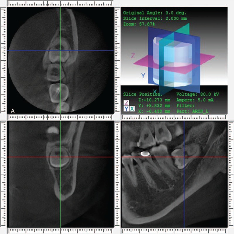
FIGURE 1.

Cone-beam computed tomography (CBCT) image of axial view, paraxial view, and sagittal view of a lingual positioned fully impacted mandibular 3rd molar.

Cone-beam computed tomography (CBCT) image of axial view, paraxial view, and sagittal view of a lingual positioned fully impacted mandibular 3rd molar.