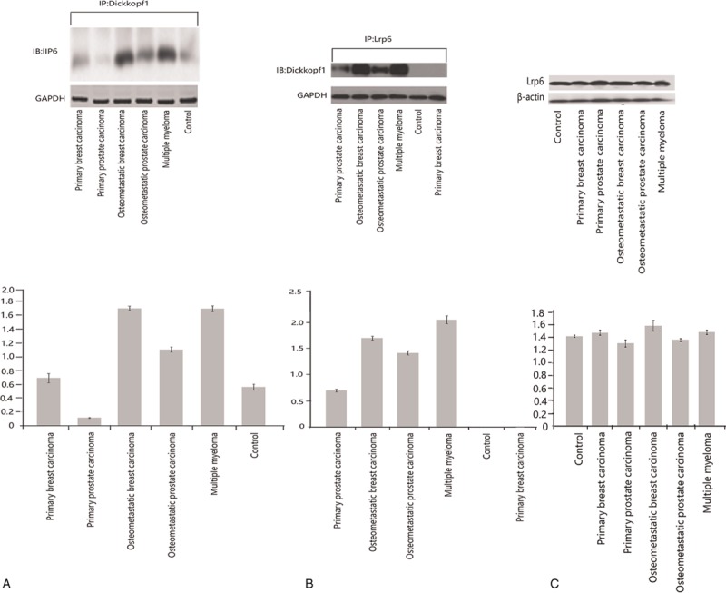
Figure 3.

(A and B) Coimmunoprecipitation of the pulled-down Dickkopf-1–Lrp6 immunoblots with either antibody shows the binding. (C) Unaltered levels of Lrp6 in the osteometastatic lesions compared with the controls. All of the panels are representative Western blots. (A and B) In the far left and middle panel, the immunoprecipitated lysates were blotted for Dickkopfl-1 and Lrp6, respectively. GAPDH was used to demonstrate equal loading of the loading lanes. The histograms below each blot are the quantitative intensities of the protein signals. (C) The far right panel shows the expression of Lrp6 in the control, primary, and metastatic lysates. β-actin was used as a loading control. The histogram below represents the quantified intensities of the protein signals. All of the assays were performed in triplicate from pooled samples. GAPDH = glyceraldehyde-3-phosphate dehydrogenase.
