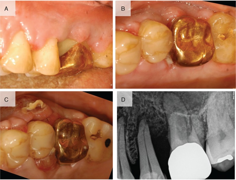
Figure 4.

(A) Clinical photograph at 2 weeks after surgery showing uneventful healing. (B) Occlusal view at 2 weeks after surgery. (C) Relapse of the lesions in the hyperplastic and neoplastic tissue was noted 4 weeks after the surgery. (D) Periapical radiograph showed bone loss between the maxillary left second premolar and the maxillary left first molar.
