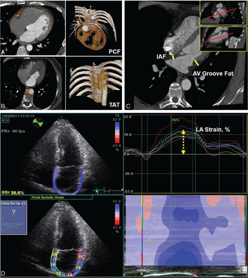
Figure 1.

(A, B) Measurements of total volume of peri-cardial fat tissue (PCF) and thoracic periaortic fat tissue (TAT). Orange color indicated PCF and TAT in axial, sagital, coronal views, and 3-dimensional reconstructions. (C) Thickness of PCF in interatrial septum (solid line) and left atrioventricular groove (dotted line) was measured in the horizontal long-axis view. (D) Left atrial (LA) deformation (LA strain, %) analysis by using 2-dimensional speckle-tracking technique and corresponding curves were displayed.
