
Figure 2.
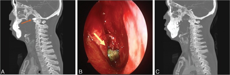
The second case: (A) sagittal CT scan showed that the bullet was located at the cranial ridge junction region; (B) intraoperative image of the bullet under the endoscopy. (C) sagittal CT scan confirmed the removal of the bullet (the arrows show the location of the bullet). CT = computed tomography.