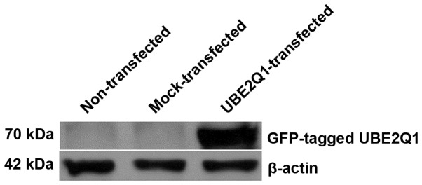
Figure 1.
Western blot analysis of UBE2Q1 protein. The right lane shows a ~70 kDa protein band representing green fluorescent protein-tagged UBE2Q1 protein. No intense band was observed for mock-transfected or non-transfected cells. Anti-β-actin antibody was used as an internal control. UBE2Q1, ubiquitin-conjugating enzyme E2 Q1; GFP, green fluorescent protein.

