
FIGURE 2.
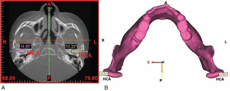
Measurements of the HCA: (A) on the horizontal CBCT image; (B) in the 3D model. 3D = three-dimensional; A = the anterior direction; CBCT = cone-beam computed tomography; HCA = horizontal condylar angle; L = the left direction; P = the posterior direction; R = the right direction.