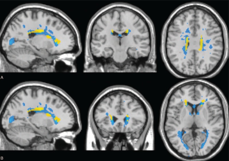
FIGURE 1.

Voxel-based analysis results obtained using fractional anisotropy maps and mean lesion mask in radiologically isolated syndrome (RIS) patients. Saggital, coronal, and axial views are presented. Clusters of reduced fractional anisotropy in RIS patients compared with healthy controls (P < 0.001) are shown in yellow and average lesion mask is shown in blue. The overlay of the significant map clusters on the mean lesion mask shows that most of the abnormalities highlighted by voxel-based analysis were primary located within lesions. (A) Both cynculate gyri. (B) Bilateral frontal sub-gyral regions.
