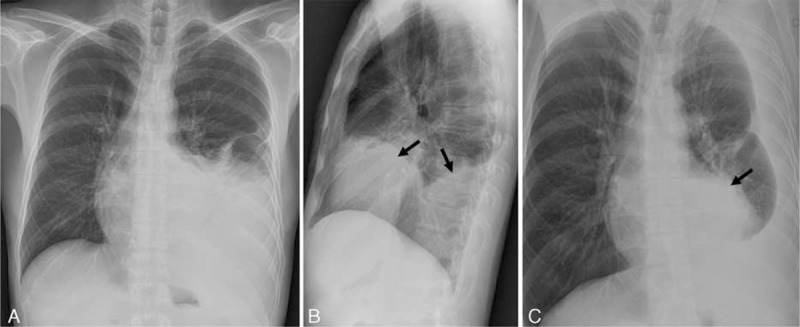
FIGURE 1.

Initial chest radiographs on admission in a 47-year-old man. Chest posterior anterior (A), left lateral (B), and left decubitus (C) views show moderate amount of left pleural effusion. Left lateral (B) and left decubitus (C) views reveal anterior and posterior mediastinal contour bulging mass opacities (black arrows in B and C), which are obscured on chest posterior anterior view (A).
