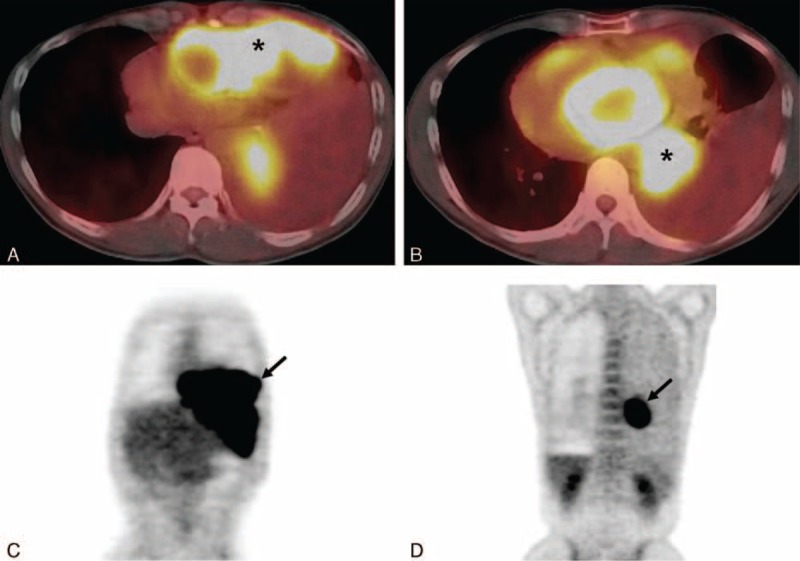
FIGURE 3.

Positron emission tomography-computed tomography scan in a 47-year-old man. Axial fusion images (A, B) show increased fluorodeoxyglucose (FDG) uptakes (maximum standardized uptake value (SUVmax, 8.3) in the anterior and posterior mediastinal masses (each asterisk in A, B). Torso maximum intensity projection images (C, D) reveal increased FDG uptake in each of the anterior (arrow in C) and posterior (arrow in D) mediastinal masses. FDG = fluorodeoxyglucose.
