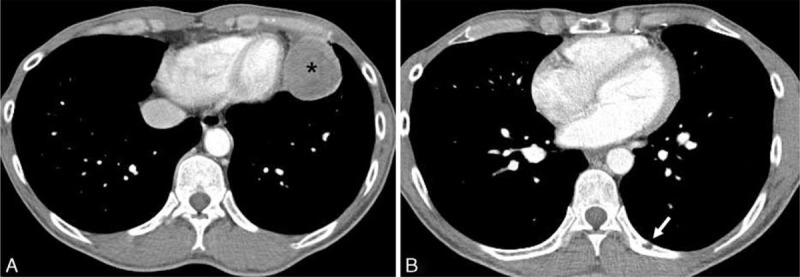
FIGURE 6.

Follow-up contrast-enhanced chest CT scan 4 months after the mediastinal tumor excision in a 47-year-old man. Axial contrast-enhanced chest CT scan performed on postoperative 4-month follow-up shows a recurrent heterogeneously enhancing left anterior mediastinal mass (asterisk) in the left paracardiac area (A). A newly appeared focal osteolytic metastatic lesion (arrow) is noted on posterior arc of the left 9th rib (B). CT = computed tomography.
