
FIGURE 3.
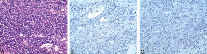
Pathology revealed that the tumor cells were uniform in size, markedly heteromorphic with eosinophilic cytoplasm, and formed nests and cords containing prominent blood sinuses (A) (HE × 200). Immunohistochemical staining was positive for hypoxia-inducible factor-1α (HIF-1α) (B) and p53 (C) (EliVision, ×200).