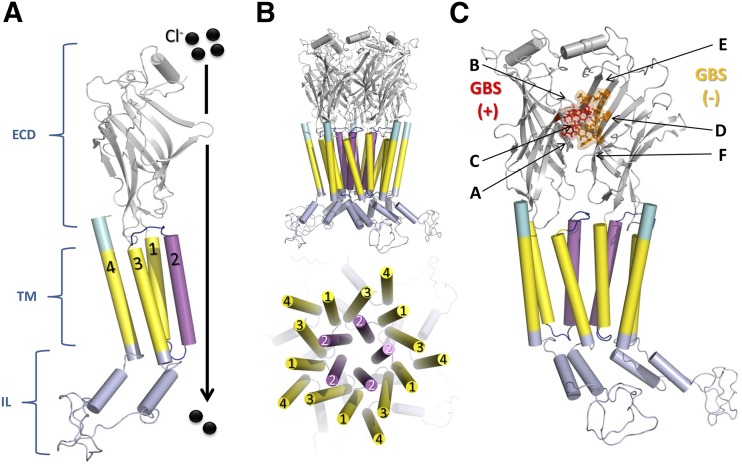
Fig. 1.
Topology and schematic structure of GlyR. (A) Representation of a monomer of α1 GlyR where the different regions of the receptor are presented: extracellular domain (ECD, gray), transmembrane domains (TM, yellow) highlighting the TM2 which is part of the channel pore (magenta), the intracellular loop domain (IL, light blue), and C-terminal region (cyan). (B) Pentameric arrangement of subunits to form functional GlyR (C) Representation of dimer α1–α1 GlyR where the glycine binding site (GBS) is located. The loops (A–C) and β-strands (D–F) that comprise the GBS are also labeled. The amino acids from the principal (F44, F63, R65, L117, L127, S129) and complementary subunit (F158, Y202, T204, F207) are colored red and orange, respectively. The IL is based on the α1 GlyR full model described previously by Burgos et al. (2015a). All images were created using PyMOL.

