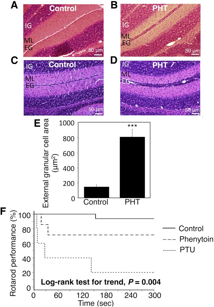
Fig. 3.
Effect of exposure to phenytoin during the perinatal period on migration of granule cells and rotarod performance in hUGT1 mice. Dams and their pups were treated with phenytoin daily from gestation day 12, 13, or 14 to postnatal day 0 (p.o.) and from postnatal day 1 to day 14 (s.c.). On postnatal days 14, the midsagittal sections of the cerebellum vermis in control mice (A and C) and phenytoin (PHT)-treated mice (B and D) were stained with hematoxylin and eosin. (E) External granule cell area was measured within 50 μm in the horizontal direction of the external granule cell layer from randomly selected fields (n = 6). Prior to the rotarod study, control mice, phenytoin-treated mice, and PTU-induced hypothyroidism mice were trained on a rotarod at a fixed speed of 6 rpm for 5 minutes. In the rotarod study, the speed of the rotarod was accelerated from 6 to 20 over 5 minutes. The time to fall off the rod was measured. (F) The difference in rotarod performance between control mice (solid line, n = 15), phenytoin-treated mice (dashed line, n = 7), and PTU-treated mice (dotted line, n = 5) was analyzed by the Kaplan-Meier method and the log-rank test for trend. Error bars show the S.D. of biologic replicates. ***P < 0.001 compared with control. EG, external granule layer; IG, internal granule layer; ML, molecular layer.

