FIGURE 2.

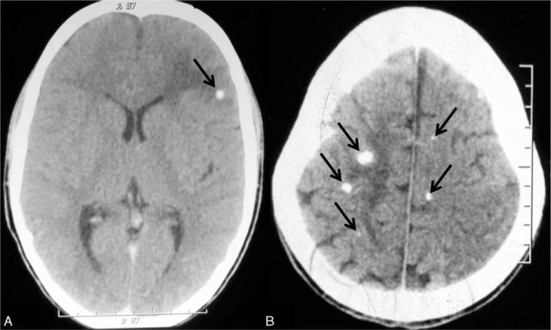
Noncontrast computed tomography of the brain showing single calcified neurocysticercosis with perilesional edema (A, arrow) and multiple calcified neurocysticercosis with perilesional edema (B, arrows).

Noncontrast computed tomography of the brain showing single calcified neurocysticercosis with perilesional edema (A, arrow) and multiple calcified neurocysticercosis with perilesional edema (B, arrows).