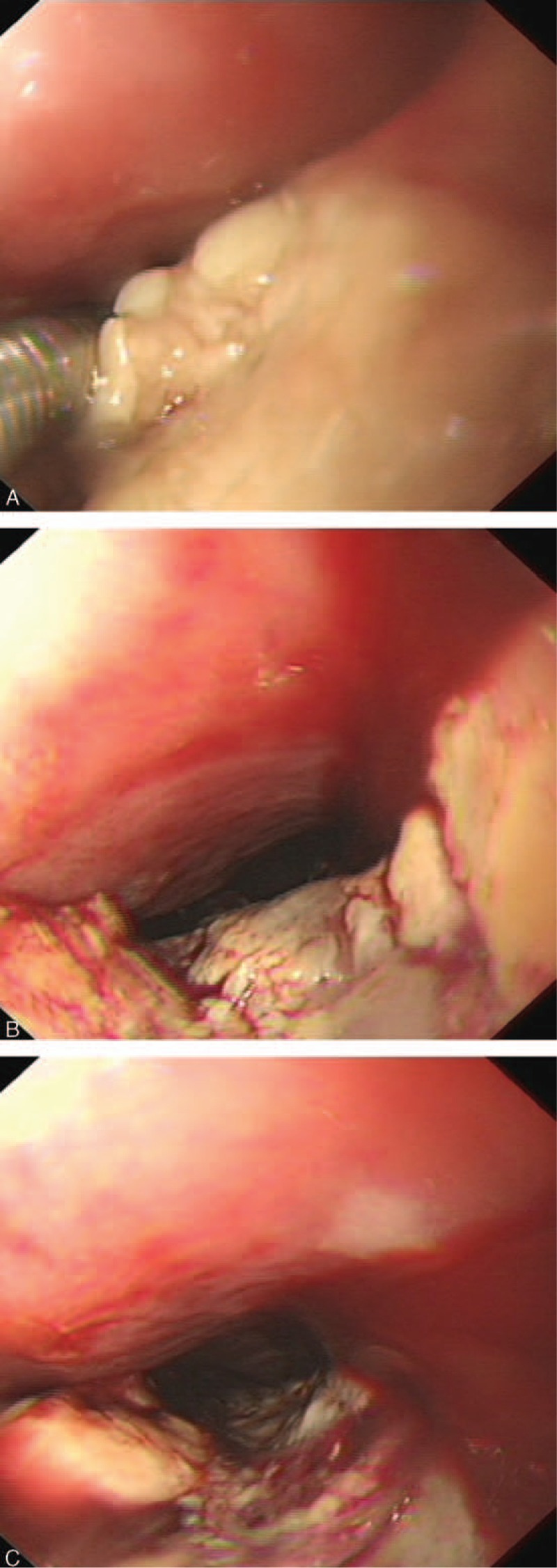
FIGURE 1.

A–C presented the endoscopic images of the primary malignant melanoma of the esophagus from different perspectives. Figure 1 A indicated that there was no pigmentation on the surface. Figure 1 B and C presented that the mass extended progressively for 15 cm along the esophageal longitudinal axis and invaded half of the esophageal circumference. Figure 1 C indicated that the mass had extensive necrotic tissue, active hemorrhage spots and no pigmentation on the surface.
