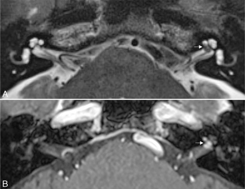
FIGURE 2.

Intralabyrinthine schwannoma. (A) An axial T2-weighted image shows a hypointense filling defect in the basal turn of the left cochlea and fundus of the internal auditory canal (arrow). (B) An axial contrast-enhanced T1-weighted image demonstrates an enhancing mass in the basal turn of the left cochlea with extension into the fundus of the internal auditory canal (arrow).
