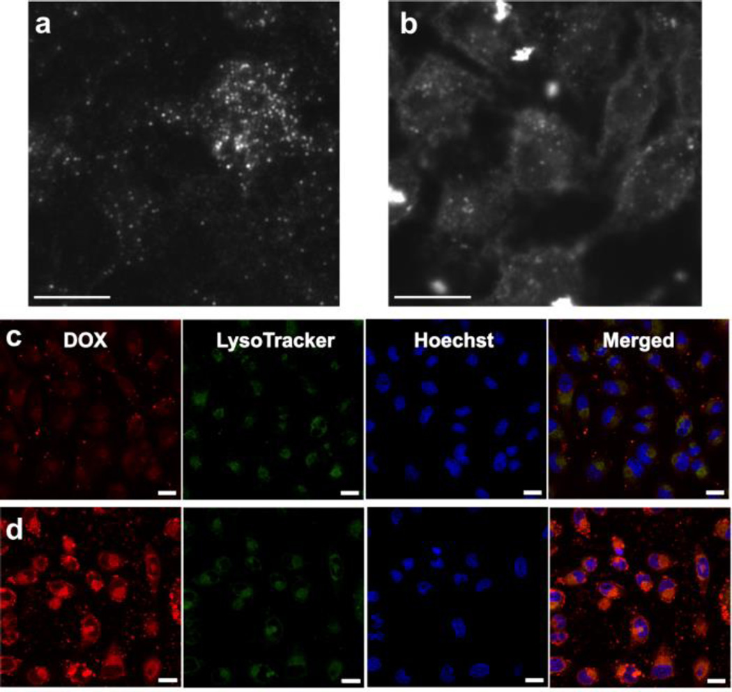
Figure 3.
In vitro studies using HeLa cells. Dark-field images of HeLa cells incubated with AuNR-DexAzo complex for 4.0 h at 37 °C: A) without FA targeting ligand and B) with FA targeting ligand (Scale bars: 10 µm). Intracellular delivery of DOX in HeLa cells was observed using confocal laser scanning microscope (CLSM) after incubation with DOX-loaded FA-CD-AuNR-DexAzo at 37 °C for C) 0.5 h and D) 4.0 h (Scale bars: 20 µm). The cells were stained by LysoTracker Green solution for 30 min and Hoechst 33342 solution for 10 min prior to CLSM imaging.

