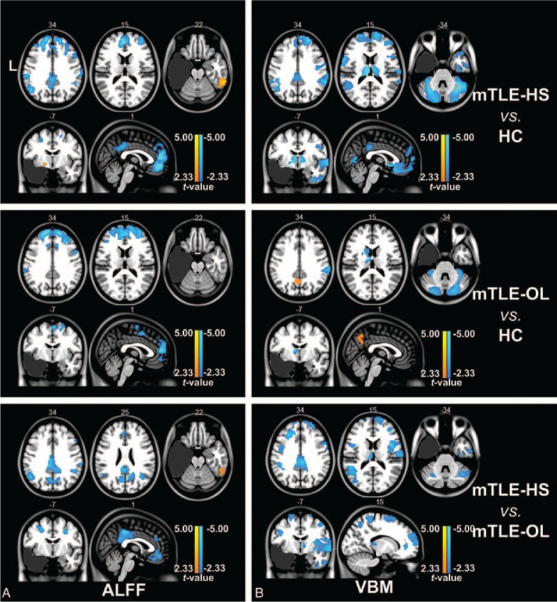
FIGURE 1.

Two-sample t tests under 1-way ANOVA of imaging parameters between groups. (A) Group comparison of ALFF between mTLE patients and healthy controls using 2-sample t test (P < 0.01, AlphaSim correction). Both mTLE-HS and mTLE-OL patients showed decreased ALFF (labeled in cold color) in bilateral anterior cingulate cortex and frontal gyrus compared with HC group. However, mTLE-HS showed decreased ALFF in bilateral frontal cortex, inferior parietal gyrus, and posterior cingulate cortex areas compared with mTLE-OL. (B) Group comparison of GMV between mTLE patients and healthy controls using 2-sample t test (P < 0.01, AlphaSim correction). MTLE-HS and mTLE-OL showed different distribution patterns of abnormalities of GMV compared to HC. In comparison with the mTLE-OL patients, decreased GMV was distributed in contralateral insula, mesial prefrontal lobe, bilateral cingulate cortex, and precuneus in mTLE-HS. Abbreviations: ALFF = amplitude of low-frequency fluctuation, HC = human controls, HS = hippocampalsclerosis, L = left, mTLE = mesial temporal lobe epilepsy, OL = other lesions, VBM = voxel-based morphometry.
