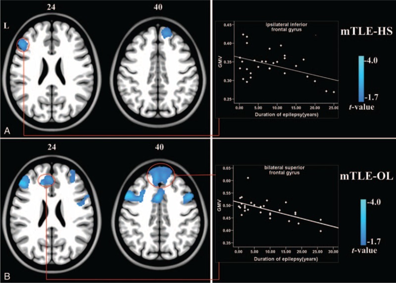
FIGURE 2.

Correlation analyses between epilepsy duration and GMV of mTLE patients. (A) The frontal lobe areas in mTLE-HS patients displayed negative correlation between GMV and epilepsy durations. (B) The bilateral superior frontal gyrus in mTLE-OL patients showed negative correlation between GMV and durations. There results were thresholded at P < 0.05 (Alphsim correction). The right scatter plot gave an example of correlation between epilepsy duration and GMV values within the cluster marked by circle. Abbreviations: HS = hippocampal sclerosis, L = left, mTLE = mesial temporal lobe epilepsy, OL = other lesions.
