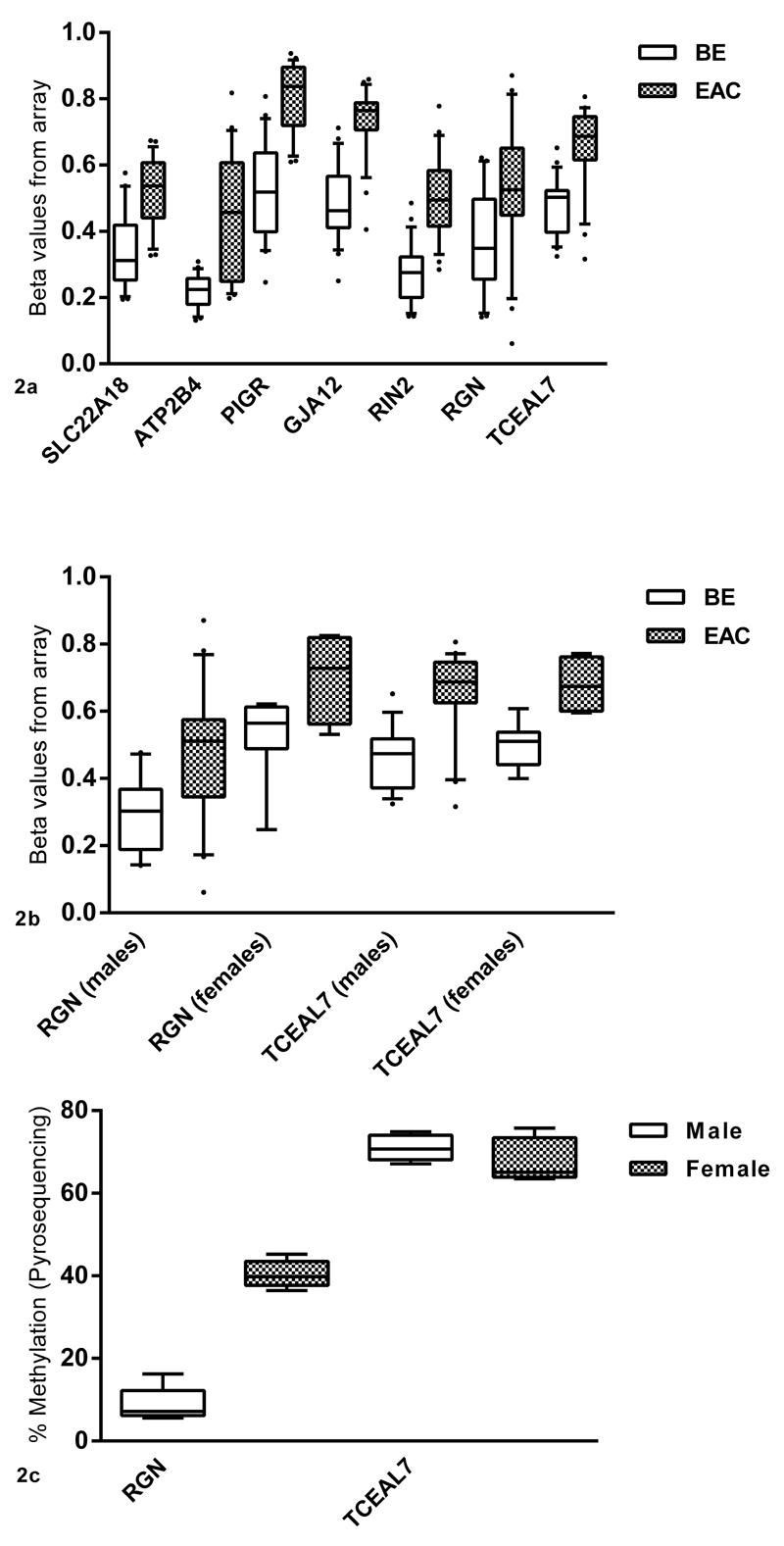
Figure 2.
a: Genes selected from the array analysis showing the greatest difference in methylation between BE and EAC. Beta values from the array are plotted on the x-axis against the gene name and tissue type on y-axis. b: For genes on the X-chromosome, analyses were separated on the basis of gender to cater for the effects of X-inactivation in females. Since RGN lies on the region of X-chromosome that is inactivated, males and females have different levels of methylation. Females have higher methylation in both tissues (BE and EAC) compared to males. TCEAL7 does not appear to be affected by X-inactivation and males and females have similar levels of methylation in both BE and EAC. c: Methylation levels for RGN and TCEAL7 in the normal esophageal epithelium in males and females using pyrosequencing (N=5).

