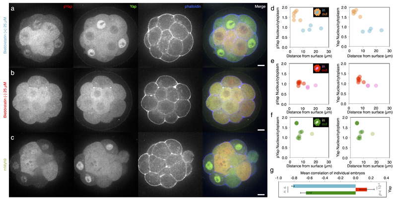
Extended Data Figure 6. Contractility controls Yap sub-cellular localization.
(a-c) Immunostaining of wild-type embryos treated with 25 µM Bb(+) (a, an inactive enantiomere of the inhibitor) or Bb(-) (b, the selective inhibitor of myosin II ATPase activity) for 3 h or mMyh9 embryos (c) showing Yap (green), phosphorylated Yap (pYap, red) and phalloidin (blue).
(d-f) Nucleus to cytoplasm intensity ratio of pYap (left) and Yap (right) as a function of the distance from the surface for WT embryo treated with 25 µM Bb(+) (d, outer cells in orange and inner cells in blue, corresponding embryo shown in a) or Bb(-) (e, outer cells in magenta and inner cells in red, corresponding embryo shown in b) or mMyh9 embryo (f, outer cells in dark green and inner cells in light green, corresponding embryo shown in c).
(g) Mean ± SEM Pearson correlation values between the nucleus to cytoplasm intensity ratio of Yap as a function of the distance from the surface from individual embryos. 252 cells from 29 embryos for Bb(+), 201 cells from 26 embryos for Bb(-) and 132 cells from 12 embryos for mMyh9 from 3 experiments each. Student t test p value is shown, n. s. for not significant.

