Fig. 1.
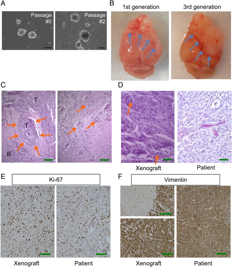
Pathological characterization of patient-derived malignant meningioma model MN3. (A) Microscopic pictures of MN3 tumorspheres cultured at passages 0 and 2. Scale bars, 100 µm. (B) Macroscopic appearance of lethal subdurally transplanted MN3 tumors located at the frontal region of mouse brains. Left, first generation. Right, third generation. Arrows point to brain-tumor boundaries. (C) H&E staining of MN3 xenografts showing whorl formation by tumor cells (left, arrows) and irregular tongue-like protrusions indicative of invasion to the brain (right, arrows). T, tumor; B, brain. Scale bars, 200 µm. (D) Higher magnification pictures of H&E-stained MN3 xenografts (left) and patient tumor sections (right), showing histological similarities between the two. Arrows show mitoses. (E and F) Immunohistochemistry for Ki67 (MIB-1) and vimentin, showing high proliferative activity and mesenchymal protein expression, respectively, in the matched patient (right) and xenograft (left). Left panels in (F) show a tumor–brain interface with negative staining in the brain parenchyma on the left (upper panel) and homogeneous staining at the tumor core (lower panel). Scale bars in (D)–(F), 100 µm.

