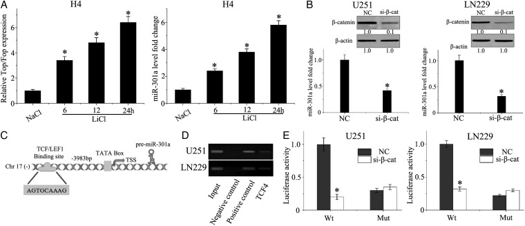
Fig. 2.
miR-301a is activated by the Wnt/β-catenin pathway by direct binding to the promoter region. (A) TOP/FOP-FLASH and quantitative PCR were used to detect Wnt/β-catenin activity and miR-301a expression in H4 cells incubated with LiCl or NaCl at 20 mM for 24 hours. (B) LN229 and U251 cells were transfected with β-catenin siRNA or scrambled siRNA using lipofectamine 2000. The expression of β-catenin and miR-301a was detected by Western blot and qPCR, respectively. (C) TCF/LEF1 binding to the promoter region of miR-301a. (D) Predicted TCF4 binding site in the promoter region of miR-301a was validated by chromatin immunoprecipitation (ChIP) PCR. The negative control was incubated with nonimmune IgG, and the positive control was incubated with anti-RNA polymerase II). (E) The pGL3-WT-miR-301a-promoter-Luc reporter or pGL3-MUT-miR-301a-promoter-Luc reporter was transfected into LN229 or U251 cells, which were then treated with β-catenin siRNA. *P < .01 as compared with the control group.

